Diagram Of Chest Area : Anatomy Of The Breast Memorial Sloan Kettering Cancer Center
Get link
Facebook
X
Pinterest
Email
Other Apps
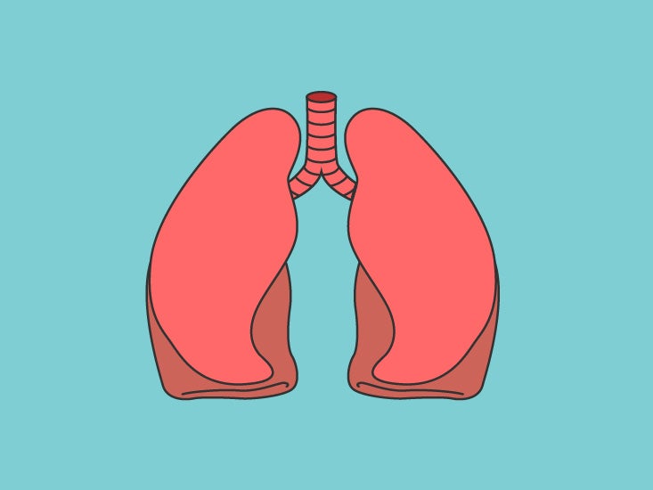
Diagram Of Chest Area : Anatomy Of The Breast Memorial Sloan Kettering Cancer Center. That's all of the acupuncture points that are a located on the chest area of the human body. See exquisite, precious, and luxurious treasure chest locations, chest map, chest locations, respawn times, chest routes & chest farming route. The chest is the area of origin for many of the body's systems as it houses organs such as the heart, esophagus, trachea, lungs, and thoracic diaphragm. A woman's chest — like the rest of her body — is covered with skin that has two layers. This page provides an overview of the chest muscle group.
This is the area from the bottom end of the neck to the top of the front legs. Your pectoralis major and pectoralis minor muscles make up most of the muscle mass in your chest. Most hernias don't need treatment, but some people eventually need surgery. It is a flat bone about six inches in length, around an inch wide, and only a fraction of an. The hindquarters encompass the he large muscular area of the hind legs and are located above the stifle and behind the barrel.
Chest Organs Anatomy Diagram Function Body Maps from post.healthline.com Angina is the term for chest pain caused by poor blood flow to the heart. That's all of the acupuncture points that are a located on the chest area of the human body. The chest anatomy includes the pectoralis major, pectoralis minor and the serratus anterior. Your pectoralis major and pectoralis minor muscles make up most of the muscle mass in your chest. The nervous system of the thorax is a vital part of the nervous system as a whole, as it includes the spinal cord, peripheral nerves, and autonomic ganglia that communicate with and control many vital organs. The abdominal cavity is the part of the body that houses the stomach, liver, pancreas, kidneys, gallbladder, spleen, and the large and small intestines.the diaphragm marks the top of the abdomen and the horizontal line at the level of the top of the pelvis marks the bottom. The chest is the area of origin for many of the body's systems as it houses organs such as the heart, esophagus, trachea, lungs, and thoracic diaphragm. The ribs and sternum make up what is called the 'ribcage.' the ribcage protects the lungs, blood vessels, and heart.
The diaphragm, a sheet of muscle in the middle chest area, is essential for breathing.
Find out more about the individual muscles within the chest anatomy by clicking their respective links throughout this Nerves of the chest and upper back. This pericardium is attached to the diaphragm, spinal column and other parts via strong ligaments. See exquisite, precious, and luxurious treasure chest locations, chest map, chest locations, respawn times, chest routes & chest farming route. See chest anatomy stock video clips. The diaphragm, a sheet of muscle in the middle chest area, is essential for breathing. The heart is enclosed in the pericardium which is a double layer. They make up the lateral part of our body, its anterior and posterior wall and they entirely build the lateral parts of the chest wall. Another of the minor extraordinary vessels, the yin linking vessel features a couple points on the chest area. Costochondritis, sometimes called costosternal syndrome or anterior chest wall syndrome, merely indicates pain and tenderness in the costochondral junction, which is the area along the sides of the breastbone where the ribs attach. The nervous system of the thorax is a vital part of the nervous system as a whole, as it includes the spinal cord, peripheral nerves, and autonomic ganglia that communicate with and control many vital organs. There isn't a specific armpit muscle, so the strained muscle causing your pain is likely your pectoral, triceps or latissmus dorsi. The abdominal cavity is the part of the body that houses the stomach, liver, pancreas, kidneys, gallbladder, spleen, and the large and small intestines.the diaphragm marks the top of the abdomen and the horizontal line at the level of the top of the pelvis marks the bottom.
Sensory information from the body and critical signals. The heart is enclosed in the pericardium which is a double layer. It lies between the right and left lungs, in the middle of the chest and slightly towards the left of the breastbone. The nervous system of the thorax is a vital part of the nervous system as a whole, as it includes the spinal cord, peripheral nerves, and autonomic ganglia that communicate with and control many vital organs. Anatomy of the chest and shoulder, anatomy of the chest organs, anatomy of the chest wall, anatomy of the chest wall and pleura, anatomy of upper chest area, human.
Heart Picture Image On Medicinenet Com from images.medicinenet.com Nerves of the chest and upper back. The nervous system of the thorax is a vital part of the nervous system as a whole, as it includes the spinal cord, peripheral nerves, and autonomic ganglia that communicate with and control many vital organs. Diagram of the emergence of the bronchial arteries in the descending thoracic aorta. Most hernias don't need treatment, but some people eventually need surgery. This is the area from the bottom end of the neck to the top of the front legs. Another of the minor extraordinary vessels, the yin linking vessel features a couple points on the chest area. See chest anatomy stock video clips. Sensory information from the body and critical signals.
This page provides an overview of the chest muscle group.
Connective tissue called the mesentery holds the abdominal organs together. Most hernias don't need treatment, but some people eventually need surgery. See chest anatomy stock video clips. It also has branching energy pathways all over the chest. They make up the lateral part of our body, its anterior and posterior wall and they entirely build the lateral parts of the chest wall. Chest pain has many possible causes, all of which need medical attention. Pectoralis major trigger point diagram, pain patterns and related medical symptoms. Find out more about the individual muscles within the chest anatomy by clicking their respective links throughout this The anatomy of the human ribs (costae) are one of the integral parts of the chest wall; Anatomy of the chest and shoulder, anatomy of the chest organs, anatomy of the chest wall, anatomy of the chest wall and pleura, anatomy of upper chest area, human. Diagram of the emergence of the bronchial arteries in the descending thoracic aorta. A woman's chest — like the rest of her body — is covered with skin that has two layers. Possible causes of pain include trauma, musculoskeletal.
Location of chest pain during angina or heart attack diagram in this image, you will find an upper chest, substernal radiating to neck and jaw, substernal raiding down left arm, substernal radiating down left arm, epigastric radiating to neck, jaw, and arms, neck and jaw, left shoulder and down both arms, intrascapular in it. Your pectoralis major and pectoralis minor muscles make up most of the muscle mass in your chest. The hindquarters encompass the he large muscular area of the hind legs and are located above the stifle and behind the barrel. Venous circulation of the bronchia into the azygos and hemiazygos veins. Most hernias don't need treatment, but some people eventually need surgery.
Cg Image Of Woman S Chest Area Heart Major Arteries And Veins Stock Images Page Everypixel from media.istockphoto.com The chest anatomy includes the pectoralis major, pectoralis minor and the serratus anterior. It lies between the right and left lungs, in the middle of the chest and slightly towards the left of the breastbone. Pectoralis major trigger point diagram, pain patterns and related medical symptoms. This pericardium is attached to the diaphragm, spinal column and other parts via strong ligaments. See exquisite, precious, and luxurious treasure chest locations, chest map, chest locations, respawn times, chest routes & chest farming route. Any diaphragm pain can, therefore, be very alarming. The chest is the area of origin for many of the body's systems as it houses organs such as the heart, esophagus, trachea, lungs, and thoracic diaphragm. The major muscle in the chest is the pectoralis major.
Most hernias don't need treatment, but some people eventually need surgery.
Your heart is surrounded by important blood vessels and arteries which pump blood into and out of your heart. Chest pain is a symptom, and so is stomach or esophagus pain, bloating, belching, and a sour taste in back of your throat. It lies between the right and left lungs, in the middle of the chest and slightly towards the left of the breastbone. It is a flat bone about six inches in length, around an inch wide, and only a fraction of an. Chest pain has many possible causes, all of which need medical attention. Sensory information from the body and critical signals. The ribs and sternum make up what is called the 'ribcage.' the ribcage protects the lungs, blood vessels, and heart. Another of the minor extraordinary vessels, the yin linking vessel features a couple points on the chest area. Any diaphragm pain can, therefore, be very alarming. The heart is enclosed in the pericardium which is a double layer. This pericardium is attached to the diaphragm, spinal column and other parts via strong ligaments. They make up the lateral part of our body, its anterior and posterior wall and they entirely build the lateral parts of the chest wall. The anatomy of the human ribs (costae) are one of the integral parts of the chest wall;
Chemal Gegg- Linda / CHEMAL-GEGG LINDA - SET 32 - 35P | Free hot girl pics . Torrent downloads » other » chemal & gegg linda set 080. March 12, 2021 april 22, 2021. 375.1 mb | total pics: You can stop your search and come to the tor search engine. By dd | posted in chemal & gegg, ichan, rachel model, sets |. Linda • chemal & gegg → amf • all models forum. You can stop your search and come to the tor search engine. Chemal gegg alissa model sets 1 112. I love models forum › teen modeling agencies › models foto and video archive chemal & gegg mega pack chemalgegg models. Discover more posts about chemal. CHEMAL-GEGG SARAH MODEL - SET 201 - 51P | Free hot girl pics from img.loveygirl.cc Discover more posts about chemal. 375.1 mb | total pics: Linda • chemal & gegg → amf • all models forum. #russia #chemal #flowers #river #mountains #tr...
Anime Gif Wallpaper Hd - Gif Wallpapers Group 62 . The best gifs for anime wallpaper. Free anime live / animated wallpapers. Log in to save gifs you like, get a customized gif feed, or follow interesting gif creators. In our group you will find the best animated artworks and anime wallpaper for dessert. Share the best gifs now >>>. Сортировать по самые популярные за неделю. Search, discover and share your favorite anime wallpaper gifs. Share the best gifs now >>>. Available 108 hight quality live wallpapers, hd animated wallpapers. A collection of the top 46 anime live wallpapers and backgrounds available for download for free. 11 Download Wallpaper Anime Gif Hd Sachi Wallpaper from media2.giphy.com Share the best gifs now >>>. We hope you enjoy our growing collection of hd images to use as a. Follow the vibe and change your...


Comments
Post a Comment